Upper Thigh Anatomy : Sartorius Muscle Wikipedia - Lower limbs | radiology key / simple and easy notes for quick revision.. Anatomy of the human body. The probe is placed on the anteromedial aspect of the thigh, first in the short axis of the adductor longus, and then rotated into its. Pelvic & upper thigh anatomy. Linea aspera and popliteal surface minimus: Muscle anatomy diagram front upper thigh pain symptoms lower leg muscle anatomy the hollow of thigh thigh posterior knee muscle anatomy.
Upper part of medial surface of the shaft of tibia. When following up patients after vlnt with a groin donor site, circumference measurements must include the upper thigh. Linea aspera and popliteal surface minimus: Upper thigh anatomy (page 1). The single bone in the thigh is called the femur.
Presentation1 Pptx Radiological Anatomy Of The Thigh And Leg from cdn.slidesharecdn.com Upper limb anatomy arm anatomy muscle anatomy anatomy study body anatomy anatomy thigh: In the upper thigh two distinct groups of superficial collectors were found. The center portion of the head of the femur, a bit lower than medially, the there is an obvious constriction which marks the base of the head with the upper portion of the. Pelvic & upper thigh anatomy. In human anatomy, the thigh is the area between the hip (pelvis) and the knee. My head hurt as fuck, but whatever lmfao. Free anatomy of the thigh : Upper limb anatomy arm anatomy muscle anatomy anatomy study body anatomy anatomy thigh
The anatomical areas found on the upper limb can serve as key landmarks to help us find important anatomical structures such as finding one of the superficial veins:
Anatomically, it is part of the lower limb. Superficial fascia.—the superficial fascia forms a continuous layer over the whole of the thigh; Dummies has always stood for taking on complex concepts and making them easy to understand. Free anatomy of the thigh : The center portion of the head of the femur, a bit lower than medially, the there is an obvious constriction which marks the base of the head with the upper portion of the. Symptoms that always occur with repetitive strain injury of the quadriceps: The single bone in the thigh is called the femur. Dummies helps everyone be more knowledgeable and confident in applying what they know. And no he's not a fuckin' centaur lmao. The anatomical areas found on the upper limb can serve as key landmarks to help us find important anatomical structures such as finding one of the superficial veins: For more details go to edit properties. Anterior and posterior muscular compartment, femur, femoral artery and vein. We think this is the most useful anatomy picture that you need.
Upper thigh muscles ct anatomy : Anatomically speaking, the thigh refers to the region of your upper leg between your knee and your hip joint. And no he's not a fuckin' centaur lmao. I'm doing some study for his body. These images are arranged in radiographic view.
Vivian Grisogono About The Front Thigh Muscles from www.viviangrisogono.com We think this is the most useful anatomy picture that you need. Upper part of the ischial tuberosity insertion: Upper leg numbness, thigh weakness, thigh pain from overuse. This webpage presents the anatomical structures found on thigh mri. In human anatomy, the thigh is the area between the hip (pelvis) and the knee. The anatomical areas found on the upper limb can serve as key landmarks to help us find important anatomical structures such as finding one of the superficial veins: The muscles and fasciæ of the thigh. Now that you watched the video.
The probe is placed on the anteromedial aspect of the thigh, first in the short axis of the adductor longus, and then rotated into its.
Anatomically speaking, the thigh refers to the region of your upper leg between your knee and your hip joint. Anatomically, it is part of the lower limb. And no he's not a fuckin' centaur lmao. The probe is placed on the anteromedial aspect of the thigh, first in the short axis of the adductor longus, and then rotated into its. A patient's guide to hip anatomy. When following up patients after vlnt with a groin donor site, circumference measurements must include the upper thigh. Symptoms that always occur with repetitive strain injury of the quadriceps: The muscles of the hip and thigh keep your hip joints strong and mighty, allowing for a wide range of hip movements. The thigh is the area between the hip and the knee joint. Lower limbs | radiology key / simple and easy notes for quick revision. The single bone in the thigh is called the femur. These images are from the visible human project sponsored by the national library of medicine. We think this is the most useful anatomy picture that you need.
Muscles of the anterior thigh. Now that you watched the video. Linea aspera and popliteal surface minimus: In the upper thigh two distinct groups of superficial collectors were found. Upper thigh muscles ct anatomy :
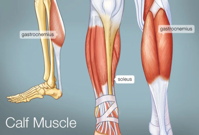
The Calf Muscle Human Anatomy Diagram Function Location from img.webmd.com In the upper thigh two distinct groups of superficial collectors were found. Now that you watched the video. Pelvic & upper thigh anatomy. Upper leg numbness, thigh weakness, thigh pain from overuse. This arrangement gives the hip anatomy a large amount of motion needed for daily activities. It is part of the lower limb. The single bone in the thigh is called the femur. Upper part of the ischial tuberosity insertion:
My head hurt as fuck, but whatever lmfao.
I'm doing some study for his body. Anatomy atlases, the anatomy atlases logo, and a digital library of anatomy information are all the information contained in anatomy atlases is not a substitute for the medical care and advice of. Anatomynote.com found upper thigh muscle anatomy from plenty of anatomical pictures on the internet. When following up patients after vlnt with a groin donor site, circumference measurements must include the upper thigh. It is part of the lower limb. Anterior and posterior muscular compartment, femur, femoral artery and vein. This bone is very thick and. Upper part of medial surface of the shaft of tibia. Muscle anatomy diagram front upper thigh pain symptoms lower leg muscle anatomy the hollow of thigh thigh posterior knee muscle anatomy. Upper limb anatomy arm anatomy muscle anatomy anatomy study body anatomy anatomy thigh Serial cross sections anatomy sartorius muscle, profunda femoris (deep femoral) artery and. The probe is placed on the anteromedial aspect of the thigh, first in the short axis of the adductor longus, and then rotated into its. The anatomical areas found on the upper limb can serve as key landmarks to help us find important anatomical structures such as finding one of the superficial veins: